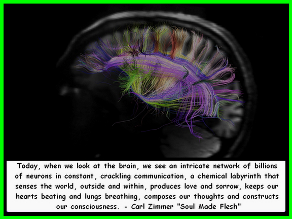
“Because science has long taught us to rely on what we can see and touch, we often don’t notice that our spirit, thoughts, emotions, and body are all made of energy. Everything is vibrating. In fact, each of us has a personal vibration that communicates who we are to the world and helps shape our reality.” – Penny Pierce Frequency: The Power of Personal Vibration
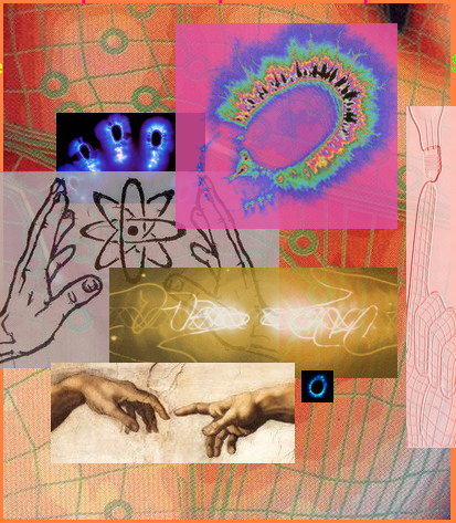
Introduction: My interest in learning why the features in the hands could be interpreted to accurately describe a person’s unique individuality led me to studying the science and biology behind the connections between our mind, body and soul. In this more than a decades search I found that our biology and our sense of self can be found inside of each other. Our hands are wired to sense and interact with our environment and our entire being. This interaction is reflected in a matrix of fields and patterns.
In the report below I have attempted to bring together the various biological and neurological connections and impulses that relate to our individuality and illustrate how these are directly connected to the hands. In brief it shows evidence as to why we can read behavior and personality in our hands. My interest in the triradii of the dermatoglyphics as human impulse markers became the initial theme for this report but the research took on a life of its own as you will see as you read on.
This report was presented at the IBMBS Biometrics Conference in Thailand in the summer of 2011.

Star–Delta Triradii: Human Impulse Markers
Patti (Lightflower) Leibold (© 2011)
We come into this world with our own unique set of life path instructions marked in our hands. Forensic science has shown that the chance of duplicate fingerprints is 1 in 64 billion.1 Continuously evolving research provides us with a wealth of data illustrating the close relationship in the developmental timing of the primary systems and organs of the body, and the hand.
Human hands contain mechanisms to transmit details sensed from the environment outside the body as well as to receive information from within the body. The hands are fascinating instruments, for in their development their individually unique communication system is permanently encoded on the glabrous skin or ridged surface of the palms.
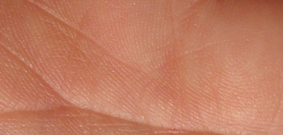
The object of this presentation is to show that we have ample evidence from research to illustrate the strong relationship between the arrangement of the papillary ridges, the creases and the triradii which divide the fields in hands, and the manner in which people sense and perceive themselves and their environment. Think about how individual just one fingerprint is, and then the greater concept: the unique patterning of the entire palm as well as the fingers. The combinations are endless!
Early researchers studied the palmar and plantar surfaces and compared the different formations to the needs of the creature. The more intelligent and evolved the species, the more complex the patterns found.2
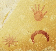
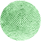
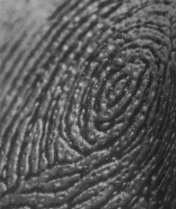
The entire palmar surface is covered with what is called glabrous skin. This skin is covered with fields of parallel rows of ridges. The distal pads of the fingertips contain patterns that range from the simple wave of ridges flowing horizontally across the fingertip to double or triple spirals spinning ‘clockwise and counter clockwise’3 around the fingertip pad.
It is interesting that the palm is covered with rows of ridges and furrows much like the brain. The bumps in the brain are called sulci and the grooves are called gyri. This folding in the brain allows for a greatly increased amount of cerebral cortex to fit inside the skull. It is quite possible that, as in the brain, the lengthening of the ridge strands formed by curving and folding back into patterns allows for increased function in small territories of the palm.

According to one source, the surface of the brain is 324 square inches: a full page of a newspaper, if spread out.4 Our palms would have a greater surface, too, if the ridges were stretched out flat. As in the palms, there are common arrangements of the patterns, but no two are alike.
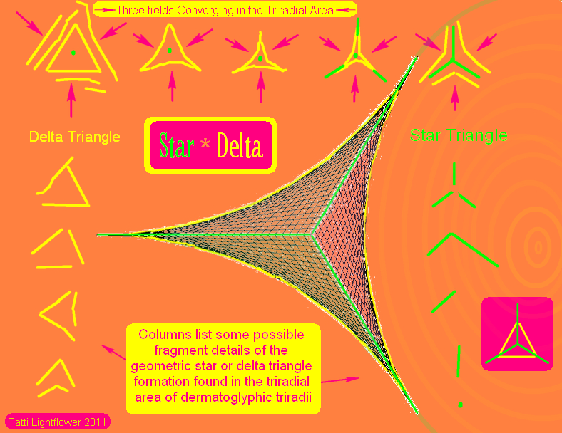
The fields of ridges are separated by triradii and the radiants that can be traced from them. In animals, where the paw pads meet on the palmar surface, a triradius is formed in the crevice between the pads. This is described as either a triangular or a pointed star shape, having three sides spaced nearly equally apart. In primates, deep creases form at the lowest level between the pads and can be traced from the triradii around the pads. Human palms are flatter, so the main creases do not directly follow the main lines from the triradii, although they are often close.5
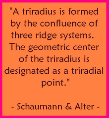
Cummins & Midlo made note of these triradii, but described them as secondary to the patterns. Technically, triradii in the human hand represent the boundaries where three different fields meet at a common location.
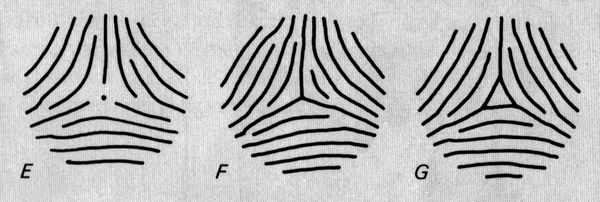
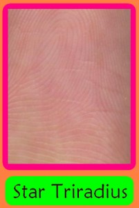
Penrose narrowed down triradii formations to two basic types.6 One type is formed by a ridge from each of the three fields meeting at the center to form a three pronged star. The other type is formed by the innermost ridge from each of the three fields meeting to form a triangular pattern with closed corners. This is called a delta formation. Many variations can be found in the spectrum between the Star and Delta Triradii.
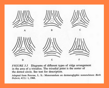
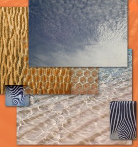
Dermatoglyphics is the term for the scientific study of fingerprints and the papillary or epidermal ridges on the palms. Cummins & Midlo compared the waves of ridges to waves of wind blown sand and cloud formations.5 Vibration from music will cause sand placed on top of a plate connected to the sound source to form into patterns, patterns which change as the music is changed.
The ridged skin of the fingertips contains far more nerve endings than the grooves or furrows that lie between the ridges. A decrease in nerves is also true of the main flexion creases. The ridges lean slightly; this leaning is called imbrication of the papillary ridges. It is because of this leaning that if you stroke a fingertip across a textured surface in one direction the surface feels rough, but if stroked in the opposite direction feels smoother.7
Typically, the distal tip of the finger has ridges that slant toward the tip. Other sections of the fingertip ridges and the palm tilt in other directions. The more complex a pattern, the more multi-directional will be this tilting. A whorl pattern could be seen as a spiral cone of multi directional sensors. An arch is the opposite of a whorl pattern. Statistics show that when there is more of one type there is less of the other. A loop is the middle pattern in the transition of the whorl to an arch, or an arch to a whorl. A whorl involves more than one triradii; a loop has one triradius, and an arch does not have a triradius unless it is what is called a tented arch. A fingerprint that consists of more than one type of pattern combined is classified as accidental and most likely has two or more triradii.
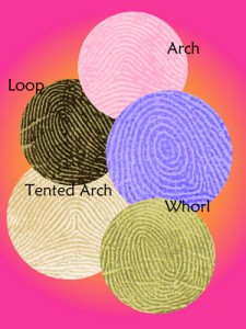
Most loops are ulnar, indicating the curving ridges around the core are located on the thumb side; others, called radial, curve in the opposite direction. Radial loops on the index finger are found in about a fourth of the population, changing sensitivity between thumb and index finger due to imbrication: the triradius of a radial loop typically would not directly contact the ridges of the thumb.
In 1823, Jan Evangelista Purkinje became the first to categorize fingerprints. He divided them into nine different types ranging from the simple arch to complicated whorls, each type transitioning to the next.8 Galton later narrowed Purkinje’s nine types to the three categories of patterns mentioned previously. Purkinje is primarily remembered as a pioneer of neuroscience. He was fascinated by vision and human comprehension. Purkinje cells in the brain and fibers in the heart are named for him and his early research.
Purkinje’s Vision, The Dawning of Neuroscience, by Nicholas J. Wade and Josef Brozek, opens with this quote:9 “It is an imperative belief of the natural scientist that each and every modification of a subjective state in the sphere of the senses corresponds to an objective state.”

Purkinje compared the fingerprints and other anatomical features to perception. Cummins & Kennedy translated Purkinje’s paper from Latin to English; which was republished in 1991 in Chris Plato’s Dermatoglyphics, Science in Transition. Purkinje wrote “After innumerable observations, I have found nine important varieties of patterns of rugae and sulci serving for touch on the palmar surface of the terminal phalanges of the fingers.”
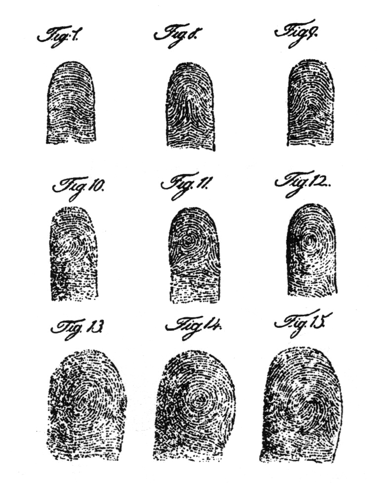
In a footnote the translators wrote “It is thus suggested that Purkinje regards the pattern formations as agents of enhanced tactile sensibility, a position held by some later workers (in contrast to the opposing but not irreconcilable view that they serve primarily for frictional augmentation).” 8 This hints at the debates in place among researchers and scientists at that time. It was acceptable that these patterns could relate to grip and be unique enough for identification purposes. Yet the time was apparently not right for recognizing their potential for understanding human perception.
Since patterns and fields of ridges develop long before birth and are virtually unique, fingerprints (and in infants, footprints) are used for individual identification.

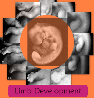
Embryological research has accelerated as technological advancements have made it possible to observe human development at earlier and earlier stages. The speed, order, and timing of the processes involved in the formation of the hands, fingers and dermatoglyphs has been observed in several fields of science, and can be compared to the development of other vital organs in the body occurring at the same time.10 Many syndromes, diseases and disorders have in their descriptions lists of combinations of hand features that are commonly found.
Anthropologists have divided the purpose of dermatoglyphics into two categories. In Palmar & Plantar Dermatoglyphics in Primates, Cummins & Midlo state: “Two chief functions have been ascribed to epidermal ridges. One concerns the increase of frictional service in locomotion and prehension, in recognition of which the areas so specialized have been designated “friction-skin” by Whipple and by Wilder. The other function is the heightening of tactile sensibility – indicated in the term “Tastlinien”. Though considerable attention has been devoted to the question as to which of these functions may be preeminent… for lack of convincing evidences we are not inclined to subordinate one to the other.”5
Susan Tucker Blackburn in Maternal, Fetal, and Neonatal Physiology: A Clinical Perspective says that in the neonate, “the skin is a sophisticated and critical organ for obtaining and receiving information about the environment”. Touch is our earliest sense. Neoceptors form around the mouth 7 weeks after gestation and the hands are sensitive to touch between 10 to 11 weeks. After 12 weeks the twitching reflex movement changes to actual movement including hand to mouth. The fingertip surfaces and the lips are two of the most sensitive areas on the body containing concentrated areas of nerve endings transmitting data back to the brain about the environment.11
The sense of touch has been studied by neurologists, physicians, and philosophers from many angles – but there is no agreement as to the means by which we comprehend the data the senses send to the brain. Some say that all senses are connected to the emotions and the emotional reaction causes an action. Others believe that the senses gather data and from pondering that data we gain experience and knowledge about the environment. Combining just these two views of a highly complex subject shows how any number of individuals can process the same information differently, due to their emotional state and their mental and physical ability to process and utilize the information.

The thumb is typically one of the first features that come to mind when we think of what sets us apart from the primates. Dorothy M. Fragaszy, a contributor in the book, The Psychobiology of the Hand, wrote: “Human hands are distinguished from those of other primates by a few distinctive features which make them particularly suitable for fine dextrous action. Included among these are relatively long thumbs, which enable us to achieve pulp-to-pulp contacts between thumb and other digits, and relatively dense direct (monosynaptic) connections between corticospinal neurons and neurons innervating the hands, enabling finer and more independent movement of the digits than other species can achieve.”12
Connolly quoted from Aristotle in his above mentioned book, “Now it is the opinion of Anacagoras that the possession of these hands is the cause of man being of all animals the most intelligent. But it is more rational to suppose that man has hands because of his superior intelligence. For the hands are instruments, and the invariable plan of nature in distributing the organs is to give to each such animal as can make use of it… we must conclude that man does not owe his superior intelligence to his hands, but his hands to his superior intelligence. For the most intelligent of animals is the one who would put the most organs to good use, and the hand is not to be looked upon as one organ but as many; for it is as it were an instrument for further instruments.”13

Walter Kidd summed up his book by saying, “use of this sense in man must have contributed greatly to his better equipment for the struggle of his life and thus, in a broad way, have been governed by a slow, remorseless process of selection. This first use of the tactile sense is of course connected only with the hand.”2
We have all heard the phrase ‘use it or lose it’ in terms of an aging body – but according to Dr. Blackburn, “use of conductive pathways may increase or alter our connections. Impulse conduction provides for functional validation of connections.” In other words, if no communication or signals are being sent or received, the pathways become less conductive or disconnected. This is happening even during prenatal development.11
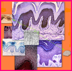
There are a number of cells that are involved in the development of the dermatoglyphic ridges. About ten weeks post-fertilization, cell proliferation begins to form shallow primary ridges at the bottom of the epidermis. These rise upward into the epidermis, becoming ridges. Cones form in the epidermis and move downward into the dermis to become sweat pores, commencing their development at about the fourteenth week. The two layers are held together like Velcro by their projections.10,14
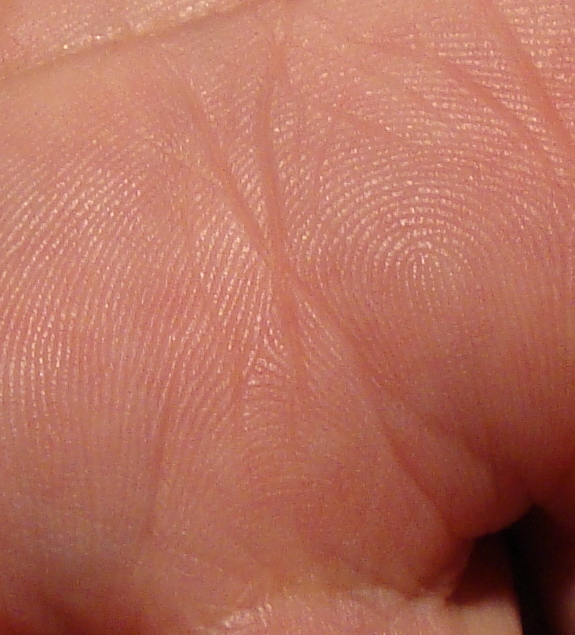
Four events precede the development of the epidermal or papillary ridge formation. These are skin innervation, Merkel cell differentiation, mesenchymal condensation and epidermal proliferation. According to researchers at the Dept. of Anatomy at Pennsylvania State University (1992) “The apical pads at the tips of the digits and the interdigital pads between the heads of the metacarpals (or metatarsals) have a unique pattern of innervation and mesenchymal content as compared to the non-pad skin. Each pad is innervated by a prominent nerve trunk and axons ascend toward the epidermis providing a density of innervation that exceeds that in the non-pad epidermis.”15
An abstract presented by V. G. Macefield from the Prince of Wales Medical Research Institute describes what is known about the sensory nerves in the periosteum. “In addition to studies on the physiology of sensory endings in human subjects, microstimulation through the recording microelectrode has revealed how the brain deals with the sensory information conveyed by a single afferent. From this work, we know that there is specificity in the sensory channels: electrical stimulation of a single Meissner or Pacinian corpuscle generates frequency dependent illusions of ‘flutter’ or ‘vibration’, whereas microstimulation of a single Merkel afferent can produce a perception of ‘pressure’ and stimulation of a single joint afferent can evoke a sensation of ‘joint rotation’.”16
Merkel cells are neuroendocrine cells that form in developing areas of the body related to patterns and to sensation such as the dermatoglyphics and hair follicles. They play a role in structure, development and function.17,18 In the book Hands the author mentioned John Hunter’s principle that “structure was the intimate expression of function”.19
Another study showed that small ridges begin to form in two sites, the center of the proximal third and the tip of the distal phalangeal or apical pad. Merkel cells were typically found in these newly forming ridges. Describing cell proliferation, the researchers concluded by saying that the dermatoglyphics can “reflect the ontogeny of the afferent nervous system that occurred prior to papillary ridge development. These observations lend support to the concept that successive waves of afferent neural development have an important role in spatial and temporal sequence of papillary ridge formation and thus the formation of both the dermatotopic map of the digits and the dermatoglyph.”20
To summarize these findings, the nervous system develops prior to ridge formation and plays a role in the formation of the ridges and their fields and patterns. Blackburn says that the earliest electrical synapses appear in the brain at 8 to 9 weeks gestation.
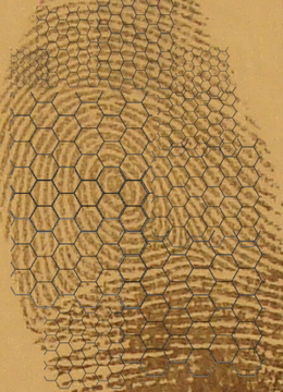
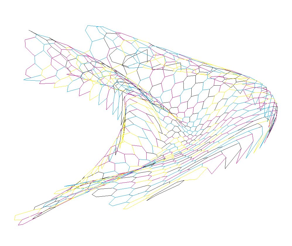
The afferent nerves developing earlier than the papillary ridges form a two-dimensional nearly hexagonal grid across the surface of the glabrous skin of the palms and fingers.21 Visualize a honeycomb grid of nerves across the entire palmar surface of the hand! Not only is there a geometric grid across the surface of the palm and fingers, but beneath the surface the vascular system along with the bundles of nerves rise from the wrists and stream across the palm in common and not so common formations.
In a paper called “Intraneural Microstimulation in Man” by Professor A. B. Vallbo, Department of Physiology, University of Umea, Sweden (1987) concluded “that the human brain has an exquisite capacity to detect, localize, delineate, and classify sensations from the input of individual tactile units in the glabrous skin of the hand. The physiological specificity of low threshold mechanoreceptors in the hand, which has been well documented in previous studies, can now be linked to distinct attributes of very simple tactile sensations which subjects report when a single afferent is stimulated electrically through an intraneural microelectrode.”22
A more recent study, in November, 2010 Alessandro Panarese and Benoni B. Edin in Italy, published their findings regarding examining “human ability to discriminate the direction of force stimuli applied to the volar surface of the index fingertip on the basis of tactile information only”. The results showed “that humans can discriminate three dimensional (3D) force stimuli whose directions differ by an angle as small as 7.1 degrees in the plane tangential to the skin surface. Moreover, we found that the discrimination ability was mainly affected by the time-varying phases of the stimulus, because adding a static plateau phase to the stimulus improved the discrimination threshold only to a limited extent.”23
Our fingers can detect the difference in temperature of only 2 degrees F and the pressure of a half a gram or the weight of a head of a pin! We can also feel certain levels of vibration in our hands even before touching the vibrating object.
Technological advancements have made it possible to validate the theories of researchers of the early 1800’s through the mid 1900’s regarding the relevance of dermatoglyphics and the sense of touch.
In the book The Sense of Touch in Animals and Birds, Walter Kidd (1907) speculated that the papillary layer of skin on the palms was primarily tactile. Referring to Meissner’s corpuscles he said: “The distribution of these touch corpuscles is significant; thus, there have been found on the ventral surface of the index-finger as many as twenty-one to the square centimeter, whereas in other parts of the palm and sole they vary from two to eight to the square centimeter.” This is in keeping with the well-known sensitivity of the pulps of the index finger. A member of his research team took a “pair of compasses, noting the distances between their points, when they were still felt as two points in such regions as the tongue and different parts of the limbs” (including the palms and sections of the fingers). The tip of the tongue was 1 mm and the distal pad of the index finger was 2 mm. Palm of the hand was 10 mm.2
A century ago Kidd also studied the areas along the flow and curvature of ridge strands where the sense of touch might be more distinct, in an attempt to learn why they lined up in rows and formed patterns.
Research today is capable of showing the reaction of only a single afferent nerve cell, and has confirmed the greater sensitivity of the index finger pad compared to the rest of the hand.


An international commission warned in “Guidelines for Limiting Exposure to Time Varying Electric Magnetic Electromagnetic Fields”, based on the results of numerous studies, that “exposure to intense pulsed microwave fields has been reported to suppress the startle response in conscious mice and evoke body movements” and “amplitude-modulated fields have also been reported to alter brain electrical activity” and leads to other health issues.26
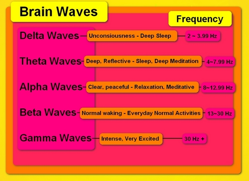
The human brain operates at various frequencies ranging from deep sleep, Delta waves at ~.5 – 4 Hz, through Theta ~4 – 8 Hz, Alpha ~ 8 – 13 Hz, Beta ~ 13 – 30 Hz, to possibly Gamma waves at ~30 – 100 Hz., which would be an intensely agitated state.
The idea that we are electrical beings isn’t new. You have probably experienced the shock from a spark of static electricity after shuffling across the floor in your slippers or been chased by a sibling finger outstretched, ready to zap you, as a child. Kirlian photography has captured the electromagnetic field around hands and living plants.
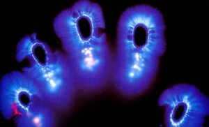
Acupuncture is a form of sending healing energy into the body using a meridian system, with needles placed in acupuncture points, and then electrically stimulated. One study reports “Many non-excitable cells have been shown electrochemical oscillation, coupling, long range intercellular communication, and can participate in meridian signal transducation.”27
Other healing methods that are based on using energy transmitted from the hands are becoming more and more accepted in the medical community. Reiki is one of these.
There is no doubt we are electrical beings with pulses and impulses.
In her book Frequency: The Power of Personal Vibration, Penney Peirce states, “Because science has long taught us to rely on what we can see and touch, we often don’t notice that our spirit, thoughts, emotions, and body are all made of energy. Everything is vibrating. In fact, each of us has a personal vibration that communicates who we are to the world and helps shape our reality.”28
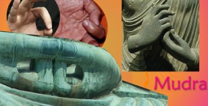
For many centuries Eastern meditations have included Mudras. Mudras are special hand positions or gestures that include touching pads of particular fingers to each other while in a relaxed meditative state of mind. Gurucharan Singh Khalsa refers to the lines and patterns in the hands, “If you understand the coding, the hands are an energy map of our consciousness and health. Each area of the hand reflexes to a certain area of the body or brain. Each area also represents different behaviors. By curling, crossing, stretching, and touching fingers to other fingers and areas we can effectively talk to the body and mind.” A mudra is a technique “for giving clear messages to the mind/body energy system”.29
“It is no coincidence that a handshake has developed as a connection between friends.” ~ Sonia Bennett Murray(34)

In summary, we are aware that electric signals are sent from cells in our hands in waves and impulses to our brains for processing. Very detailed information from the stimulation of even a single cell is made known to us.
Our hands are uniquely encoded with rows of ridges divided by triradii and their radiants. As stated earlier, triradii form (with variants) in the Star and Delta patterns, both geometric formations with near equal angles. Signals are transmitted almost instantly from nerve endings in the ridges of the palms and fingers to the brain.
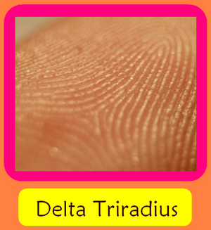
Two kinds of circuitry in electrical engineering relate to an alternating or continuously flowing current. Coincidentally, they are called Star and Delta. The Star circuitry is based on pulsing and the Delta is a steady wave of energy.
It appears that the human body operates at varying electrical frequencies and is most stressed by pulsating vibrations versus a constant wave of current. The triradii, which are the meeting locations of three different energetic, sensory fields from three different directions, may be a marker for further study into the varying levels of electrical impulse energy among humans.
It is known that the energy levels and personality types of people with few triradii are different than those with a high number of triradii.
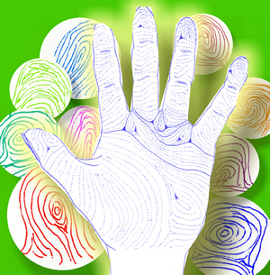
Recent studies in autism and schizophrenia show differences from non afflicted people in the way in which the senses receive information and sensory input from the environment and process it mentally and emotionally.
One study, working with twins that were discordant for schizophrenia, found that the most significant markers related to the time period between 13 and 15 weeks estimated gestational age. This is an important time period in the development of the dermatoglyphics and the continuing development of major creases in the palms.30 The proximal transverse crease – the line known as the head line in hand analysis — forms at around 14 weeks.
Another study found that certain types of schizophrenics have a number of asymmetrical features between the left and right hands, along with disturbed ridges, ridges that have broken down into dotted lines that are also called “strings of pearls”. Disturbed ridges have lost their connectivity as long strands and have broken down into individual or near individual sections of ridge islands with one or two pores.3,31, 32
These results underline the connection between the state of the dermatoglyphics and the state of mind. Further studies are needed to see if the total number and type of triradii, in Star-Delta formations, have any significance in the manner in which an individual is affected by the transmission of pulses and waves of energy.
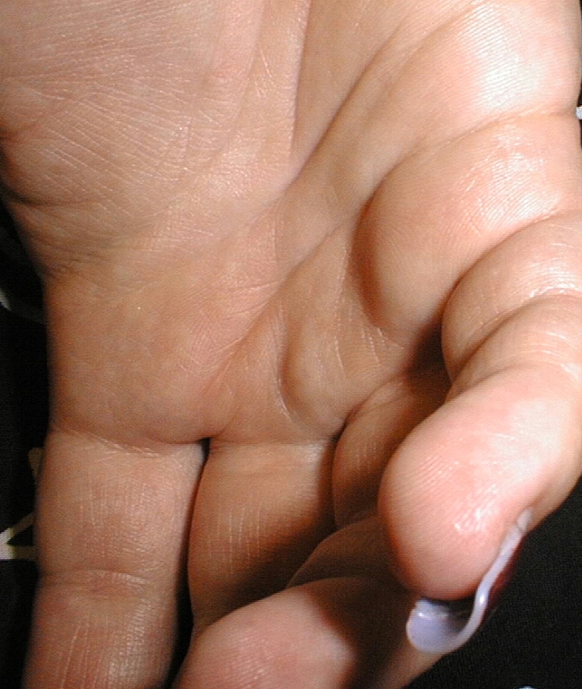
It is shown that the dermatoglyphics in their fields of parallel rows and patterns across the palmar surface are highly sensitive to sensory stimulation. These fields are divided by the main lines that can be traced from the radiants of the triradii and cut by the deeper creases transversing the palms.
Dedicated researchers have performed lengthy studies on every aspect of the hands. For centuries, hand analysts have related people’s characteristics and behaviors to features in the hands. The fields of sensitive papillary ridges and the creases that divide them form patterns and territories which can be qualitatively and quantitatively evaluated.
Ralph M. Garruto wrote in The State of Dermatoglyphics, “The field of dermatoglyphics continues to endure… The driving force behind its usefulness for anthropology, biology, genetics, and medicine can be based on the number of new investigators who use the field as a tool to solve thematic questions that are relevant to the scientific community as a whole.”33
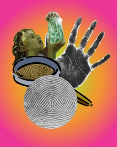
My interest in the study of behavior began with the question, “what makes us tick?” After more than three decades of studying the markers in thousands of hands as an analyst, I am certain that the fields and patterns of dermatoglyphics and the creases that cross them have a direct relationship to ‘what makes us tick.” All human hands have the same basic structure, but no two are exactly alike: even your own two hands are not mirror images of each other.
The structure, texture, flexibility and various unique markings of the human hand can be quantitatively and qualitatively studied. The intricate and extremely sensitive nerves signaling information about one’s place in the universe are visible in the patterns they form in the papillary ridges or dermatoglyphics on the surface of the palms (and feet.) Like patterns in the sand or ripples in the water, the hands contain formations that reflect the energy that moves within them.
*
References – Suggested Reading:
1Sir Francis Galton, 1892, quoted in Historical and Scientific Foundation of Friction Skin Comparison, by William Leo, 2004.
2Walter Kidd, M.D., F.Z.S., 1907, The Sense of Touch in Mammals and Birds
3Amrita Bagga, Dermatoglyphics of Schizophrenics, 1989.
4Eric H. Chudler, Faculty, Washington University
5Charles Midlo & Harold Cummins, Palmar and Plantar Dermatoglyphics in Primates, 1942; Finger Prints, Palms and Soles, 1943.
6Lionel S. Penrose, 1971, Dermatoglyphics and Medicine
7Walter Kidd, M.D., F.Z.S., 1907, “On Imbrication of the Papillary Ridges in Man”
8Harold Cummins and Rebecca Wright Kennedy, 1940, translation of Purkinje’s “Observations on Finger Prints and Other Skin Features”,(1823.) Republished in Dermatoglyphics, Science in Transition 1991
9Nicholas J. Wade and Josef Brozek, Purkinje’s Vision, The Dawning of Neuroscience, 2001
10William J. Babler, PHD, “Prenatal Communalities in Epidermal Ridge Development”, published in Trends in Dermatoglyphic Research, 1990; “Prenatal Development of Dermatoglyphic Patterns: Associations with Epidermal Ridge, Volar Pad and Bone Morphology”, 1987. “Embryologic Development of Epidermal Ridges and Their Configurations” published in Dermatoglyphics, Science in Transition 1991.
11Susan Tucker Blackburn, Maternal, Fetal, and Neonatal Physiology: A Clinical Perspective, 2007.
12Dorothy M. Fragaszy, “How Non-Human Primates Use Their Hands”, published in The Psychobiology of the Hand edited by Kevin J. Connolly 1998
13Kevin J. Connolly, The Psychobiology of the Hand, 1998
14David R. Ashbaugh, Quantitative-Qualitative Friction Ridge Analysis, 1999
15Morohunfola, Jones & Munger, Pennsylvania State University, “The Differentiation of the Skin and its Appendages, Normal Development of Papillary Ridges” 1992.
16V. G. Macefield, University of New South Wales, Sydney, Australia, “Physiological Characteristics of Low-threshold Mechanoreceptors in Joints, Muscle and Skin in Human Subjects”, 2005.
17K. Baumann, Z. Halata, I. Moll, “The Merkel Cell: Structure-Development-Function-Cancerogenesis”, 2007.
18A. Beiras, quoted from “The Merkel Cell: Structure-Development-Function-Cancerogenesis”, 2007.
19John Napier, Hands, 1993
20S. J. Moore, B. L. Munger, Pennsylvania State University, “The Early Ontogeny of the Afferent Nerves and Papillary Ridges in Human Digital Glabrous Skin”, 1989.
21D. A. Dell, B. L. Munger, “The Early Embryogenesis of Papillary (sweat duct) Ridges in Primate Glabrous Skin: The Dermatotopic Map of cutaneous Mechanoreceptors and Dermatoglyphics”, 1986.
22A. B. Vallbo, Professor, Department of Physiology, University of Umea, Sweden, 1987.
23Alessandro Panarese and Benoni B. Edin, ARTSLab, Italy, 2010 “Human Ability to Discriminate Direction of Three-Dimensional Force Stimuli Applied to the Finger Pad”
24Saal, Vijayakumar & Johansson, University of Edinburgh, UK, “Information about Complex Fingertip Parameters in Individual Human Tactile Afferent Neurons”, 2009.
25Nestor Hugo Mata, National Technical University, Electrical Engineering Department, Argentina, “What is the Level of Radio-Frequency Waves that Affect Human Health”.
26”International Commission on Non-Ionizing Radiation Protection (ICNIRP) Guidelines for Limiting Exposure to Time Varying Electric Magnetic Electromagnetic Fields (up to 300 GHz)”, 1998.
27Charles Shang, Boston School of Medicine, “The Meridian System and The Mechanism of Acupuncture”, 1996.
28Penney Peirce, Author, Counsellor, Frequency: The Power of Personal Vibration, 2009.
29M.S.S. Gurucharan Singh Khalsa, Sadhana Guidelines for Kundalini Yoga Daily Practice, 1978.
30J. O. Davis and H. S. Bracha, Department of Psychology, SW Missouri State University, USA, “Prenatal Growth Markers in Schizophrenia: A Monozygotic Co-Twin Control Study”, 1996.
31Avila, Sherr, Valentine, Blaxton, & Thaker, Maryland Psychiatric Research Center, Baltimore, MD, USA, “Neurodevelopmental Interactions Conferring Risk for Schizophrenia: A Study of Dermatoglyphic Markers in Patients and Relatives”, 2003.
32C. S. Mellor, Memorial University of Newfoundland, Canada, “Dermatoglyphic Evidence of Fluctuating Asymmetry in Schizophrenia”,1992.
33Ralph M. Garruto, Professor Anthropology and Neurosciences, The State of Dermatoglyphics, The Science of Finger and Palm Prints (Durham, Fox & Plato) 2000.
34 Sonia Bennett Murray (author) My Amazing Step-Mom and Editor-in-Chief <3
Star~Delta Triradii: Human Impulse Markers
All rights reserved – Patti (Lightflower) Leibold – 2011

